
Editor's Note: This post is a part of our Girls in STEM mentorship program. Emilie Reas, a neuroscience Ph.D. student at the University of California San Diego's Human Memory Lab studying memory retrieval with fMRI, teamed up with Vi Nguyen, a high school junior with a passion for science, to explore how the brain remembers past experiences. Their project aimed to understand real-life memories in the context of findings from Cognitive Neuroscience research. To learn more about Emilie and Vi, as well as our other STEM students and mentors, click here.
These maps show the brain regions that were more active when participants correctly recognized the studied words, than when they forgot. The brighter areas show the areas of the brain where blood flow and oxygenation is increased, which provides an estimation of brain activity.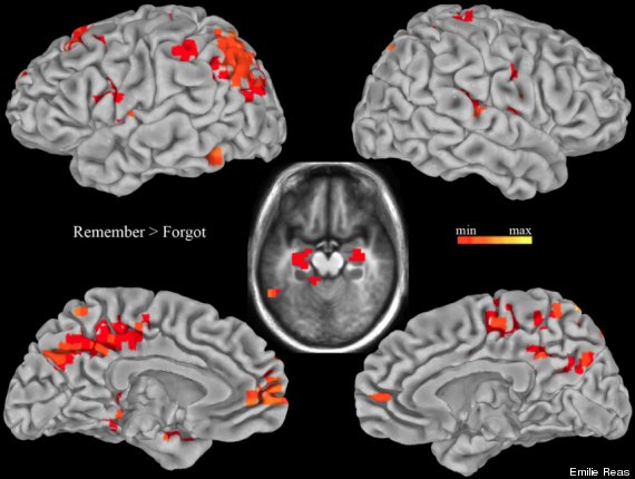
Why did you decide to work on this project?
Emilie: To integrate Vi's love for science and experiences using online social media with my graduate research.
How did you actually conduct this project?
Emilie: First, Vi surveyed users through online media, requesting respondents to share their personal memories. Next, we examined data from a recent study conducted as part of my PhD to understand how memories such as those collected from Vi's survey are studied in a laboratory setting. The study used functional magnetic resonance imaging (fMRI) while participants performed a recognition test on words they had studied either once or several times (Reas and Brewer, Hippocampus, 2013). We then looked at the fMRI data, which measures changes in blood flow and oxygenation and therefore provides an estimate for brain activity during the recognition test.
Vi: Using Microsoft Excel and Emilie's lab data, we computed the average accuracies for recognizing words studied once or several times or not at all. Then, we created graphs using Microsoft Excel to better understand the data.
What were your findings?
Vi: Using the graphs from Emilie's data, we can see that the participants most accurately recognized words that they had studied several times (about 90% of the time), while words studied only once were recognized, on average, only 64% of the time.
We also computed the average reaction times for participants to respond to the words either correctly or incorrectly, and created graphs for reaction times for words studied once or several times. The graphs show that, on average, it took participants longer to respond incorrectly than correctly. It took participants longer to remember words that they had studied just once (1.19 seconds) than words that they had studied several times (1.04 seconds).
How did you display these findings?
Vi: We used brain activation maps (above) to show which areas of the brain are most active during certain activities--in this case, memory retrieval.
Which part of the brain is most involved in memory retrieval?
Vi: The area of the brain most involved in memory retrieval is the medial temporal lobe, which is located near the bottom of the brain. The hippocampus, located in the medial temporal lobe (above in the middle image), plays an important role in the formation of memories and the retrieval of recent memories. However, it is still unclear what role the hippocampus does play. It is thought that perhaps this part helps "assembling" the memory, by taking in the various inputs from different areas of the brain (such as the visual and auditory parts of a memory) and putting them all together into one coherent memory.
What other areas of the brain also show memory activity?
Vi: It is thought that the parietal lobe's activity during memory retrieval is to assist in attention during memory retrieval, such as when looking for a memory or focusing on a memory. You can see the activity in the parietal lobe in the top left brain image--the section near the upper-back of the brain that shows brain activity.
What did you learn from this process that you did not know before?
Vi: Something new I learned about was data analysis. I have some experience with data analysis from my high school science classes, but using Microsoft Excel to quickly and easily analyze data was surprisingly fun.
What was your favorite thing about working with each other?
Vi: Working with Emilie over the past several weeks has been truly rewarding. I was able to learn more about how the brain works (a subject I find really fascinating!) as well as about how research is conducted in neuroscience. (I also discovered how much I enjoy data analysis, something I didn't expect to learn!)
Key takeaways for fellow ladies in STEM: Keep an open mind when working in new fields and experiment often.