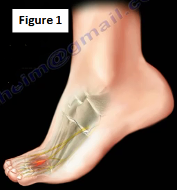
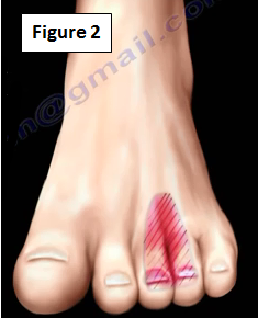
Morton's neuroma is a benign, painful condition usually affecting middle-aged women who walk in narrow shoes (Figure 1). The pain is felt in the front of the foot extending to the toes, usually in response to irritation, trauma, or excessive pressure (Figure 2).
Anatomy
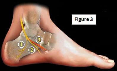
Around the tarsal tunnel area, the tibial nerve splits into three branches; the calcaneal branch (1), the lateral plantar branch (2) and the medial plantar branch (3) (Figure 3).
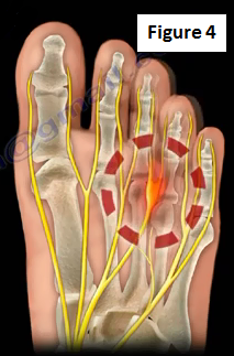
At the bottom of the foot, the medial and lateral plantar nerves give rise to the digital nerves. The neuroma is an enlargement and inflammation of a portion of the nerve. It is usually located in the third webspace between the metatarsal heads (Figure 4).
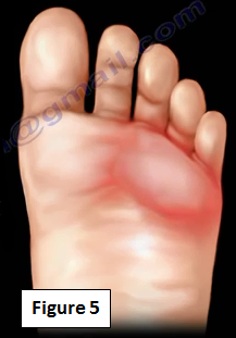
Pain from Morton's Neuroma is primarily located on the bottom of the ball of the foot (Figure 5). The pain is made worse by walking, especially while wearing narrow shows and it relieved by removing the shoe.
How do you test for Morton's Neuroma?
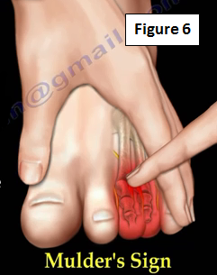
It is usually diagnosed based on clinical examinations, an MRI and ultrasound may help in the diagnosis. Examination may include a web space compression test. Squeezing the metatarsals together with one hand and using the index finger of the other hand to compress the affected the area. Applying pressure will cause pain, paresthesia, and tingling. A possible click and radiating pain into the affected toes produced the "Mulder's Sign" (Figure 6).
Ultrasound Scanning Techniques
The dorsal approach is done by placing the ultrasound on the dorsal surface and applying finger pressure in the web space from the plantar foot (Figure 7a). The plantar approach is performed by placing the ultrasound on the plantar surface and squeezing the two metatarsal heads together (Figure 7b). Applying this pressure will cause pain, paresthesia, and tingling. When this test is positive, the nerve gets squeezed and this often creates a clicking sound, and pain in the foot. Ultrasound is probably the best study. It can differentiate between a bursa which is compressible, from a neuroma which is not compressible. MRI is usually helpful.

Differential Diagnosis
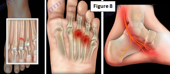
There are many different differential diagnoses associated with Morton's neuroma including metatarsal stress fracture, metatarsalgia, and lumbosacral nerve root irritation (Figure 8).
Treatment
Treatment depends on the severity of the symptoms. Therapy is a conservative treatment option where the patient will be given arch supports and foot pads. Steroid injections may also be used. Surgical release (decompression) of the intermetatarsal ligament and removal of the neuroma are considered after failure of conservative treatment.
For more information on foot pain, follow the links below:
https://www.youtube.com/watch?v=c7QewW3Up50
https://www.youtube.com/watch?v=BkGVWTeJTqs
https://www.youtube.com/watch?v=J7-L9MFRXD8
https://www.youtube.com/watch?v=KN9tz0K7l3E
https://www.youtube.com/watch?v=NrZDCGpkYro
https://www.youtube.com/watch?v=ZsvOGXxPJ7M
For more information on other topics, visit my YouTube Channel:
https://www.youtube.com/user/nabilebraheim